Frontier Papers in Spine CT AI: A Personal Review
This note summarizes the spine CT papers and technical directions I currently find most useful: vertebra segmentation, vertebral labeling, compression fracture interpretation, opportunistic osteoporosis screening, and the shift from leaderboard metrics toward deployable review pipelines.
This is a personal reading review, not clinical guidance. I am using "frontier" in an engineering sense: work that changes how I would build, test, or operate a spine CT analysis system. The most interesting papers are not always the ones with the largest model. The useful papers are the ones that clarify where errors happen, how a reviewer can inspect them, and how the output can survive a real workflow.
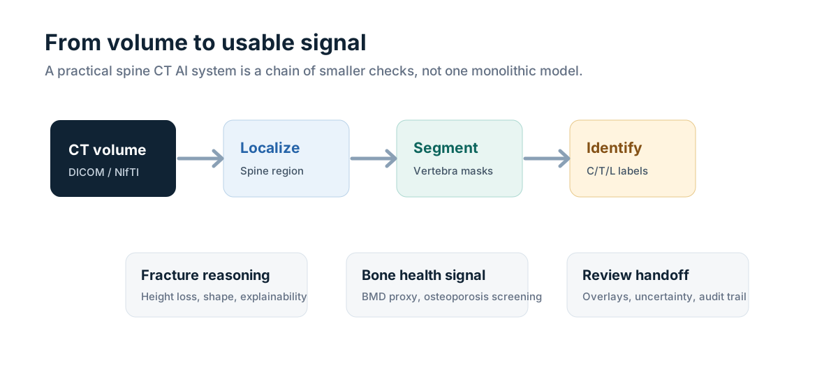
My main conclusion is simple: a practical spine CT AI system is rarely a single end-to-end model. It is a chain of smaller tasks: field-of-view detection, spine localization, vertebra segmentation, vertebral labeling, pathology-specific measurement, bone health estimation, uncertainty reporting, and human review. If one stage is unstable, the downstream "AI result" becomes hard to trust.
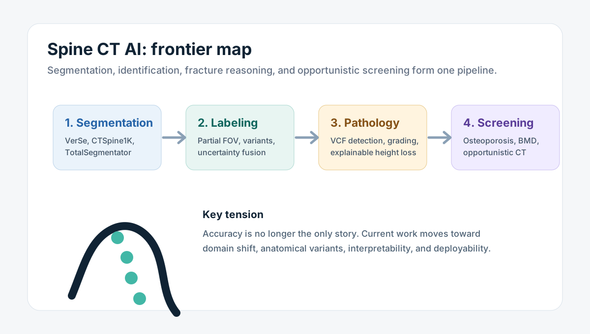
Real imaging examples used while reading
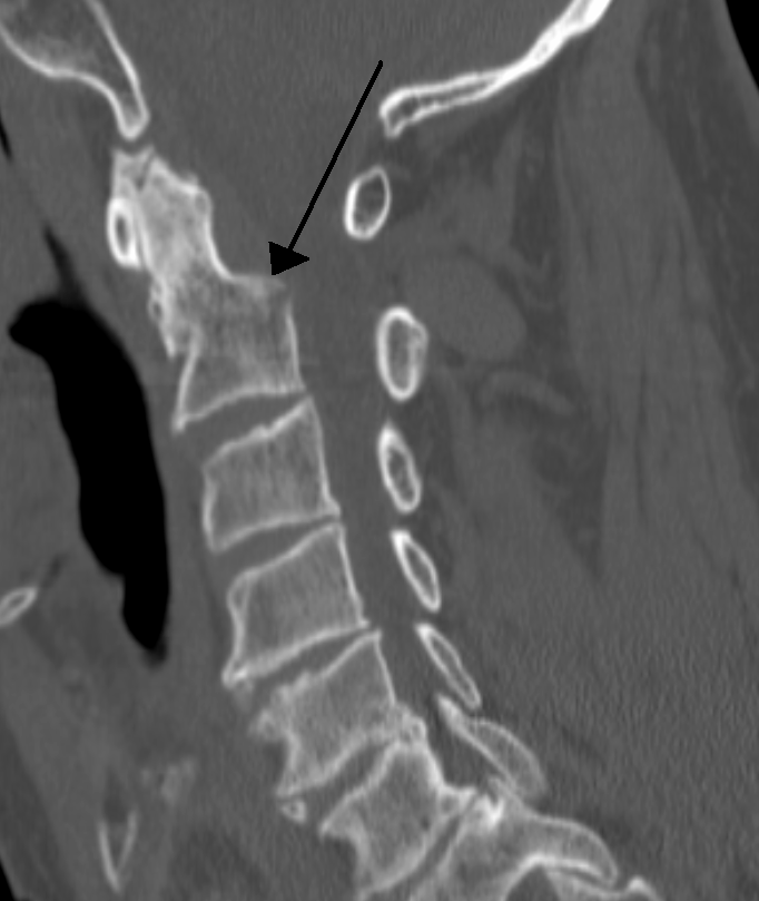
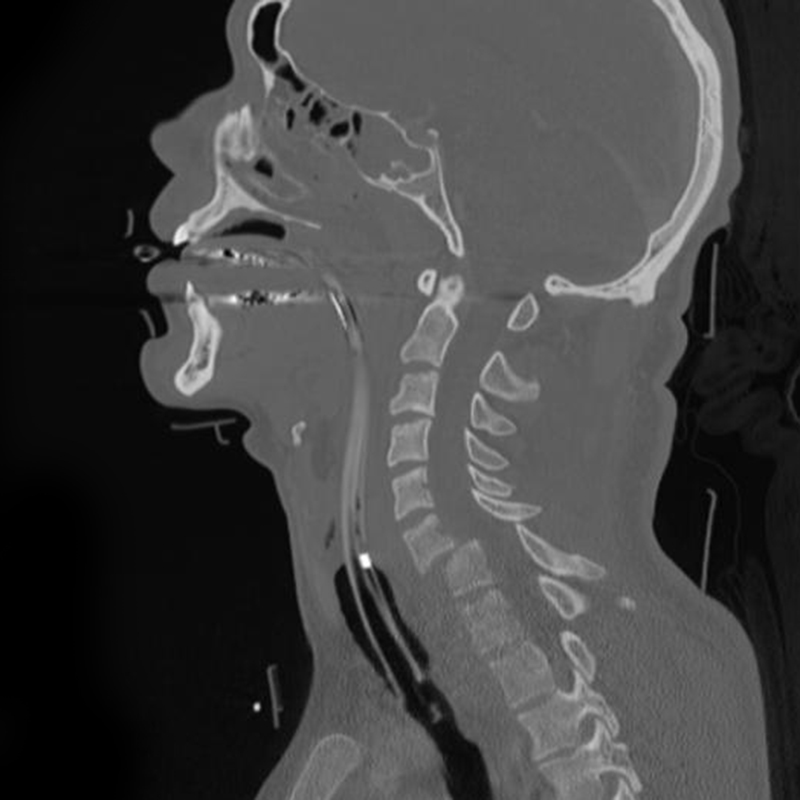
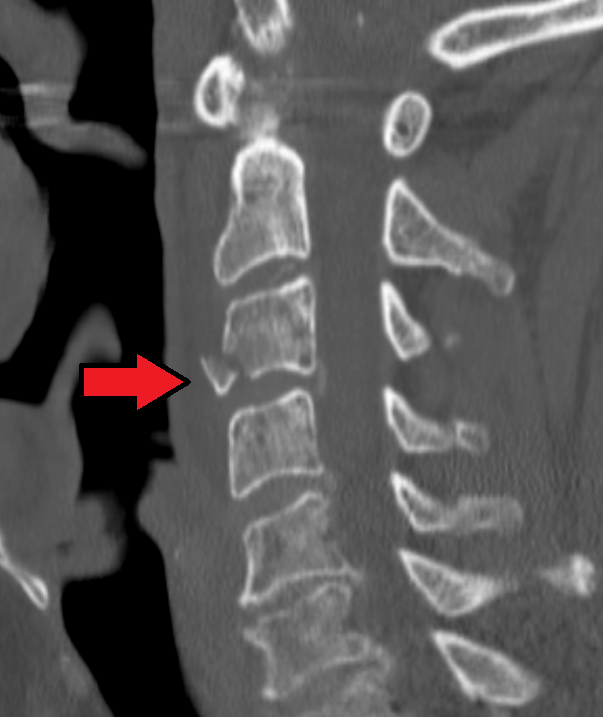
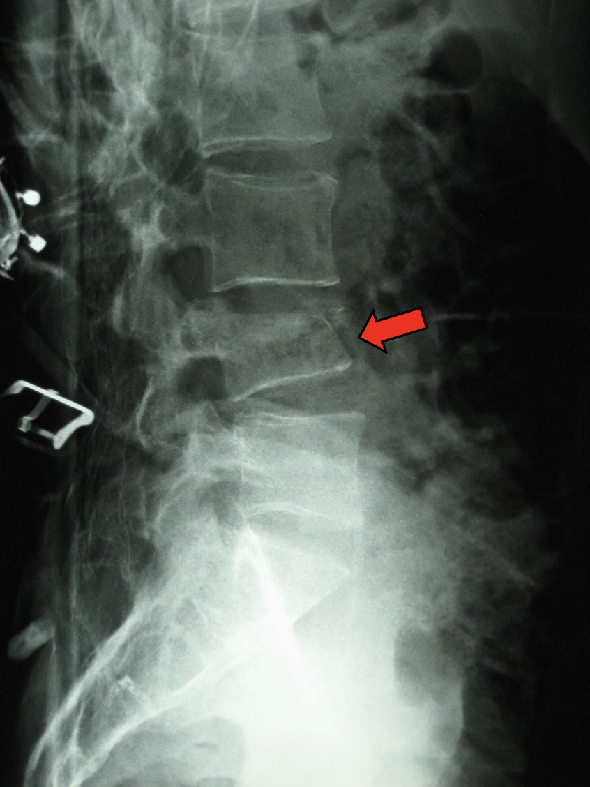
The following public images are included as visual anchors for this note. They are not cases from this site and they are not used for diagnosis here. I use them only to make the technical discussion less abstract: fracture morphology, vertebral alignment, field of view, and annotation visibility are easier to reason about with actual radiology examples on the page.




1. Why spine CT is a demanding benchmark
Spine CT looks deceptively structured. Vertebrae repeat, the anatomy is mostly rigid, and bony boundaries are often high contrast. That first impression is misleading. In real scans, the field of view may include only a few vertebrae. There may be transitional anatomy, congenital variants, severe scoliosis, degenerative change, compression deformity, tumor involvement, surgical hardware, low-dose noise, contrast variation, or partial cropping. A model can perform well on clean full-spine cases while failing on exactly the cases that matter operationally.
This is why I treat spine CT as a workflow problem before treating it as a model problem. A segmentation error may be acceptable if it is small and visible to the reviewer. A labeling error can be much more dangerous because it can move a finding to the wrong level. A fracture classifier may have a good AUC, but if it cannot explain whether the signal comes from height loss, endplate change, or acquisition artifacts, it may be difficult to integrate into review.
From that perspective, the best recent work shares a common theme: it tries to make the intermediate state explicit. Where is the spine? Which vertebra is this? How uncertain is the label? What shape cue supports the fracture decision? Is the bone density signal calibrated enough to trigger review, or is it only a weak screening hint?
2. Segmentation and labeling: the foundation layer
The VerSe benchmark remains one of the clearest starting points because it combines vertebra segmentation and vertebral labeling. That pairing is important. A mask without a correct level is not enough for clinical communication, and a level without a reliable mask is not enough for quantitative measurement. VerSe also made the community confront a practical issue: unusual anatomy and incomplete coverage are not edge cases in deployment; they are routine sources of error.
CTSpine1K extended the data side of the problem. With 1005 CT volumes and more than 11100 annotated vertebrae, it moved the field toward larger-scale training and evaluation. I see CTSpine1K as useful not just because of size, but because it encourages discussion about distribution: scanner settings, body regions, annotation consistency, and the gap between research data and operational data.
TotalSegmentator is another useful reference point. It is not a spine-only system, but that is exactly why it matters. In many engineering prototypes, a general whole-body CT segmentation model becomes the first component in a downstream pipeline. It can provide coarse anatomy, quick masks, and a standard baseline. For a personal research stack, it is often more useful to start with a robust general segmenter and then add spine-specific correction than to build every component from scratch.
The open question is how to evaluate segmentation in a way that predicts downstream utility. Dice score is useful, but it does not tell the full story. A small mask error near an endplate may affect fracture grading more than a larger error in a clinically irrelevant region. A missed posterior element may matter for some tasks and not for others. For spine CT, task-aware evaluation is likely more meaningful than a single global segmentation score.
3. Partial field of view and vertebral identity
Many routine CT scans are not spine protocols. A chest CT may include the thoracic spine. An abdominal CT may include lower thoracic and lumbar levels. A neck CT may show only cervical levels. This makes vertebral identity difficult because the model cannot always rely on seeing the full sequence from C1 to sacrum.
SpineCLUE is interesting because it focuses on vertebra identification under arbitrary field of view. Its pipeline uses localization, segmentation, and identification, with contrastive learning and uncertainty estimation to reduce confusion between visually similar vertebrae. I like this framing because it treats identity as a probabilistic, context-dependent result rather than a trivial label attached after segmentation.
In practice, I would want any deployed labeling system to expose uncertainty. If the model is unsure whether a vertebra is T12 or L1, hiding that uncertainty is worse than showing it. Downstream fracture reporting, surgical planning, and follow-up comparison all depend on level identity. A small uncertainty badge or review queue can be more useful than a falsely confident label.
4. Foundation models, SAM-style interaction, and the role of the reviewer
After the success of promptable segmentation models in natural images, it is natural to ask whether similar interaction patterns help in spine CT. SAM-style systems and vision-language interfaces are attractive because they offer a path from automatic output to user correction. A reviewer can point, box, or describe a target instead of redrawing everything manually.
SpinalSAM-R1 is representative of this direction: it combines spine CT segmentation with multimodal interaction and software-oriented workflow thinking. I would not read this kind of work as "the model replaces the operator." I read it as a sign that the field is moving from pure batch inference toward interactive correction. That matters because real imaging workflows are full of near-miss outputs: good enough to accelerate work, not good enough to accept blindly.
The deployment question is not only segmentation accuracy. It is whether the interaction is faster than manual correction, whether the user can understand failure modes, whether the system preserves an audit trail, and whether it can run inside the actual review environment. A clever prompt interface that cannot be integrated into the viewer is less useful than a simpler model that exports clean overlays and uncertainty flags.
5. Lightweight models still matter
Large models dominate attention, but lightweight 3D CNNs and efficient U-Net variants remain important. SpineContextResUNet is a useful example because it emphasizes computational efficiency and contextual modeling rather than simply increasing model scale. This is relevant for hospitals, small labs, and edge deployments where hardware is limited, data transfer is constrained, or offline processing is preferred.
I see this as a healthy counterweight to foundation-model enthusiasm. A small model with predictable memory use, fast inference, and stable behavior on the local scanner distribution may be more valuable than a larger model that is harder to validate. In medical imaging operations, maintainability is part of performance.
For my own evaluation, I would compare models along four axes: accuracy on standard benchmarks, robustness under field-of-view variation, inference cost, and ease of review integration. A model that is second-best on Dice but first-best on review latency may be the better engineering choice.
6. Vertebral compression fracture: from detection to explanation
Vertebral compression fracture detection is one of the most natural downstream tasks for spine CT AI. CT contains rich bone morphology, and many fractures are visible on scans acquired for unrelated reasons. However, a simple fracture/no-fracture classifier is not enough. Reviewers need to know why the model flagged a vertebra, whether the finding is acute or chronic, how severe the deformity is, and whether the model is reacting to a true height loss or to artifacts.
The neurosymbolic VCF work is interesting because it combines learned segmentation with rule-like reasoning over vertebral height distributions. This is a practical idea: compression fracture grading is partly geometric. If the model can show anterior, middle, and posterior height patterns, the prediction becomes easier to audit.
HealthiVert-GAN takes a different but related route by generating a pseudo-healthy vertebra and estimating relative height loss. I like the conceptual move because it reframes fracture grading as a counterfactual shape problem: what would this vertebra likely look like without compression? That does not solve every clinical ambiguity, but it provides a more interpretable measurement story than a bare probability score.
The hard part is validation. Counterfactual shape generation can look plausible while being wrong. A clinical system would need careful stress testing on degenerative deformity, scoliosis, malignancy, postoperative anatomy, and low-quality scans. Interpretability should not become decorative; it has to be tied to measurable failure modes.
7. Opportunistic osteoporosis screening
Opportunistic screening is one of the most important public-health directions for spine CT. Many patients receive chest, abdomen, trauma, or oncology CT scans that include vertebrae. Those scans can contain signals related to bone mineral density and fracture risk, even when osteoporosis was not the reason for imaging.
The value proposition is strong: use existing imaging to identify patients who may need follow-up assessment. But the technical and operational risks are also real. CT protocols vary. Contrast phase matters. Scanner calibration matters. Vertebral level matters. Degenerative sclerosis can affect apparent density. A model trained on one population or scanner distribution may not transfer cleanly.
Recent work using texture-preserving self-supervision, mixture-of-experts modeling, and multi-task integration reflects those challenges. Self-supervision is attractive because much CT data is unlabeled. Expert mixtures are attractive because the population is heterogeneous. Multi-task learning is attractive because diagnosis, BMD estimation, vertebral localization, and fracture context are related but not identical tasks.
From a systems perspective, I would not treat opportunistic osteoporosis AI as a final diagnostic system. I would treat it as a triage signal. The output should lead to review, risk stratification, or follow-up recommendation pathways, not an automatic label detached from clinical context.
8. Evaluation: what I would measure beyond Dice and AUC
The papers in this area often report Dice, identification accuracy, sensitivity, specificity, AUC, or correlation with BMD. Those are necessary, but they do not fully answer whether a model is deployable. I would add several operational metrics.
- Level-critical error rate: how often a finding is assigned to the wrong vertebral level.
- Partial-FOV robustness: performance when only cervical, thoracic, lumbar, or mixed partial regions are visible.
- Review correction time: how long it takes a human to correct the output to an acceptable state.
- Uncertainty calibration: whether uncertain labels are actually more likely to be wrong.
- Artifact resilience: behavior under metal hardware, motion, low dose, tumor, and severe deformity.
- Workflow latency: whether outputs arrive early enough to influence review rather than after the report is already complete.
These metrics are less glamorous than benchmark scores, but they are closer to what determines whether a model will be trusted in routine use.
9. My current synthesis
The spine CT field seems to be moving in two directions at the same time. One direction is broader and more interactive: foundation models, promptable segmentation, multimodal interfaces, and general anatomy models. The other direction is narrower and more operational: efficient 3D networks, explicit vertebral identity, interpretable fracture geometry, and opportunistic screening pipelines.
I do not see these directions as competitors. A mature system may use a general model for coarse anatomy, a specialized model for vertebra segmentation, a labeling module with uncertainty, a fracture measurement module, and a screening module for bone health. The user should see a coherent review product, but under the hood it will likely remain modular.
For my own study, the most important lesson is to keep intermediate outputs visible. In spine CT, the hidden errors are often worse than the obvious errors. A visibly imperfect mask can be corrected. A silently wrong vertebral label can contaminate every downstream statement.
10. Open questions I want to follow
- Can vertebral labeling models represent anatomical variants without forcing every case into a normal template?
- Can compression fracture models provide shape-based explanations that radiologists actually find useful?
- How should opportunistic osteoporosis screening handle scanner calibration and contrast-phase variation?
- Will promptable segmentation reduce correction time in real viewers, or only in demos?
- Can lightweight models reach the reliability needed for edge deployment without constant cloud inference?
- What public benchmark best predicts performance on messy hospital CT data?
References and reading list
- VerSe: A Vertebrae Labelling and Segmentation Benchmark for Multi-detector CT Images
- CTSpine1K: A Large-Scale Dataset for Spinal Vertebrae Segmentation in Computed Tomography
- TotalSegmentator: robust segmentation of 104 anatomical structures in CT images
- SpineCLUE: Automatic Vertebrae Identification Using Contrastive Learning and Uncertainty Estimation
- An Intrinsically Explainable Approach to Detecting Vertebral Compression Fractures in CT Scans via Neurosymbolic Modeling
- HealthiVert-GAN: Pseudo-Healthy Vertebral Image Synthesis for Interpretable Compression Fracture Grading
- Opportunistic Osteoporosis Diagnosis via Texture-Preserving Self-Supervision, Mixture of Experts and Multi-Task Integration
- SpinalSAM-R1: A Vision-Language Multimodal Interactive System for Spine CT Segmentation
- SpineContextResUNet: A Computationally Efficient Residual UNet for Spine CT Segmentation
- Wikimedia Commons: PdensfracCT.png
- Wikimedia Commons: Cervical fracture dislocation C6-C7
- Wikimedia Commons: TeardropFractureC3CTMarked.png
- Wikimedia Commons: L4 Compression Fracture Arrow