
Segmentation review
Human-in-the-loop mask review, confidence thresholding, and export notes for downstream reporting.
Civitas Imaging Lab is a personal technical notebook for imaging workflow engineering: static documentation, release files, validation checklists, and operational notes for small clinical AI pilots.

Each module is documented as a small, auditable workflow rather than a black-box product bundle.
Human-in-the-loop mask review, confidence thresholding, and export notes for downstream reporting.
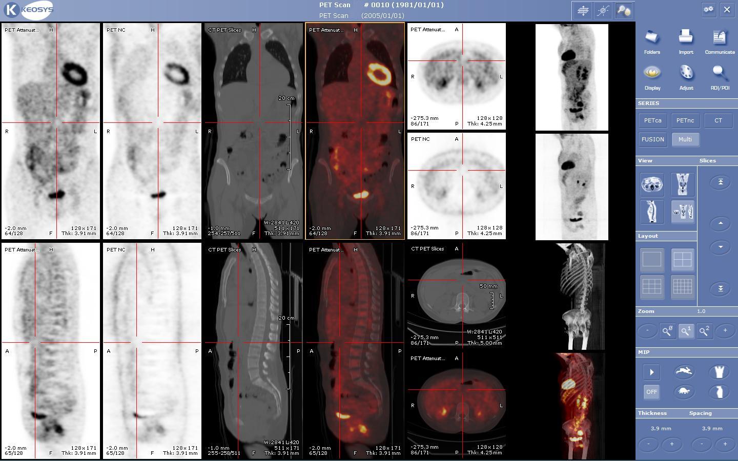
Structured queues for study prioritization, lesion bookmarks, and quality flags before radiologist review.

DICOM metadata checks, report payload mapping, and reproducible transfer manifests.
This site is written like an engineering desk reference rather than a brochure. The pages collect deployment assumptions, validation habits, release file conventions, and small operational decisions that are useful during imaging AI pilots.
| Date | Update | Area |
|---|---|---|
| 2026-06-18 | Published notes on static docs for clinical workflow pilots. | Notes |
| 2026-06-12 | Updated checksum and release file convention notes. | Downloads |
| 2026-06-04 | Refreshed status page, privacy language, and public manifest. | Operations |
Short entries about deployment hygiene, reproducible reference files, and keeping documentation useful during review.
A longer English reading note on spine CT segmentation, vertebral labeling, fracture interpretation, and opportunistic screening.
A small static documentation site can be easier to review and maintain than a full portal during early validation.